Thursday, 8 September 2011

Nucleic Acid


Nucleic acid is a macro molecule composed of chains of nonnumeric nucleotides. In biochemistry these molecules carry genetic information or form structures within cells. The most common nucleic acids are deoxyribonucleic acid and ribonucleic acid. Nucleic acids are universal in living things, as they are found in all cells and viruses. Nucleic acids were first discovered by Friedrich Miescher in 1871.Artificial nucleic acids include peptide nucleic acid, Morpholino and locked nucleic acid as well as glycol nucleic acid and threose nucleic acid. Each of these is distinguished from naturally-occurring DNA or RNA by changes to the backbone of the molecule

Types of nucleic acids: - 1.Ribonucleic acid
2. Deoxyribonucleic acid

Nucleic acid components: - 1 .Nucleo bases
2. Nucleosides
3 .Nucleotides and deoxy nucleotides.

Stranded DNA viruses


Some small viruses carry their genome as single-stranded DNA (ssDNA) molecules. These viruses have a simple genome, one gene for a viral nucleocapsid protein and another gene for a DNA replication enzyme. The virus with a ssDNA genome also faces a serious replication problem in the host cell. When introduced into cells these genomes can not be used to make viral proteins because the only template for transcription is double stranded DNA

For this reason the first step after infection is the conversion of the viral ssDNA into dsDNA using host cell DNA polymerase. You may recall that DNA polymerase requires a primer for replication. In some of these viruses the 3 end of the viral DNA folds back and forms dsDNA by base pairing with an internal sequence. In this way, the primer is built into the genome and the 3 end can be extended to create dsDNA that serves as a template for transcription. The resulting transcripts are translated to make the viral proteins the replicated viral DNA is converted back into a ssDNA genome, and the virion is packaged for export. Canine and feline parvo viruses are members of the ssDNA virus family.

Mitochondrial DNA Damage


In human cells, and eukaryotic cells in general, DNA is found in two cellular locations inside the nucleus and inside the mitochondria. Nuclear DNA (nDNA) exists as chromatin during non replicative stages of the cell cycle and is condensed into aggregate structures known as chromosomes during cell division. In either state the DNA is highly compacted and wound up around bead-like proteins called histones. Whenever a cell needs to express the genetic information encoded in its nDNA the required chromosomal region is unravelled, genes located therein are expressed, and then the region is condensed back to its resting conformation. Mitochondrial DNA (mtDNA) is located inside mitochondria organelles, exists in multiple copies, and is also tightly associated with a number of proteins to form a complex known as the nucleoid. Inside mitochondria, reactive oxygen species (ROS), or free radicals, byproducts of the constant production of adenosine triphosphate (ATP) via oxidative phosphorylation, create a highly oxidative environment that is known to damage mtDNA. A critical enzyme in counteracting the toxicity of these species is superoxide dismutase, which is present in both the mitochondria and cytoplasm of eukaryotic cells.

DNA TESTING


There have been two main types of forensic DNA testing. They are often called RFLP and PCR based testing, although these terms are not very descriptive. Generally, RFLP testing requires larger amounts of DNA and the DNA must be underrated. Crime-scene evidence that is old or that is present in small amounts is often unsuitable for RFLP testing. Warm moist conditions may accelerate DNA degradation rendering it unsuitable for RFLP in a relatively short period of time

PCR-based testing often requires less DNA than RFLP testing and the DNA may be partially degraded, more so than is the case with RFLP. However, PCR still has sample size and degradation limitations that sometimes may be under appreciated. PCR-based tests are also extremely sensitive to contaminating DNA at the crime scene and within the test laboratory. During PCR, contaminants may be amplified up to a billion times their original concentration. Contamination can influence PCR results, particularly in the absence of proper handling techniques and proper controls for contamination.PCR are less direct and somewhat more prone to error than RFLP. However, PCR has tended to replace RFLP in forensic testing primarily because PCR based tests are faster and more sensitive.

DNA Damage and Mutation


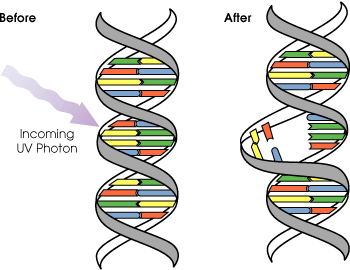
It is important to distinguish between DNA damage and mutation, the two major types of error in DNA. DNA damages and mutation are fundamentally different. Damages are physical abnormalities in the DNA, such as single and double strand breaks, 8-hydroxydeoxyguanosine residues and polycyclic aromatic hydrocarbon adducts. DNA damages can be recognized by enzymes, and thus they can be correctly repaired if redundant information, such as the undamaged sequence in the complementary DNA strand or in a homologous chromosome, is available for copying. If a cell retains DNA damage, transcription of a gene can be prevented and thus translation into a protein will also be blocked. Replication may also be blocked and/or the cell may die

In contrast to DNA damage, a mutation is a change in the base sequence of the DNA. A mutation cannot be recognized by enzymes once the base change is present in both DNA strands, and thus a mutation cannot be repaired. At the cellular level, mutations can cause alterations in protein function and regulation. Mutations are replicated when the cell replicates. In a population of cells, mutant cells will increase or decrease in frequency according to the effects of the mutation on the ability of the cell to survive and reproduce. Although distinctly different from each other, DNA damages and mutations are related because DNA damages often cause errors of DNA synthesis during replication or repair and these errors are a major source of mutation

Given these properties of DNA damage and mutation, it can be seen that DNA damages are a special problem in non-dividing or slowly dividing cells, where unprepared damages will tend to accumulate over time. On the other hand, in rapidly dividing cells, unprepared DNA damages that do not kill the cell by blocking replication will tend to cause replication errors and thus mutation. The great majority of mutations that are not neutral in their effect are deleterious to a cell’s survival. Thus, in a population of cells comprising a tissue with replicating cells, mutant cells will tend to be lost. However infrequent mutations that provide a survival advantage will tend to clonally expand at the expense of neighboring cells in the tissue. This advantage to the cell is disadvantageous to the whole organism, because such mutant cells can give rise to cancer. Thus DNA damages in frequently dividing cells, because they give rise to mutations, are a prominent cause of cancer. In contrast, DNA damages in infrequently dividing cells are likely a prominent cause of aging.

Structure of a Telomerase RNA

RNA is transcribed with only four bases (adenine, cytosine, guanine and uracil), but there are numerous modified bases and sugars in mature RNA's. Pseudouridine (Ψ) in which the linkage between uracil and ribose is changed from a C–N bond to a C–C bond and ribothymidine (T) are found in various places (most notably in the TΨC loop of tRNA). Another notable modified base is hypoxanthine, a deaminated adenine base whose nucleoside is called inosine (I). Inosine plays a key role in the wobble hypothesis of the genetic code. There are nearly 100 other naturally occurring modified nucleosides, of which pseudouridine and nucleosides with 2'-O-methylribose are the most common. The specific roles of many of these modifications in RNA are not fully understood. However it is notable that in ribosomal RNA many of the post-transcriptional modifications occur in highly functional regions, such as the peptidyl transferase center and the subunit interface implying that they are important for normal function

The functional form of single stranded RNA molecules, just like proteins, frequently requires a specific tertiary structure. The scaffold for this structure is provided by secondary structural elements which are hydrogen bonds within the molecule. This leads to several recognizable "domains" of secondary structure like hairpin loops, bulges and internal loops. Since RNA is charged, metal ions such as Mg2+ are needed to stabilize many secondary and tertiary structures.

DNA repair Mechanisms

Cells cannot function if DNA damage corrupts the integrity and accessibility of essential information in the genome (but cells remain superficially functional when so-called "non-essential" genes are missing or damaged). Depending on the type of damage inflicted on the DNA's double helical structure, a variety of repair strategies have evolved to restore lost information. If possible, cells use the unmodified complementary strand of the DNA or the sister chromatid as a template to recover the original information. Without access to a template, cells use an error-prone recovery mechanism known as translation synthesis as a last resort

Damage to DNA alters the spatial configuration of the helix and such alterations can be detected by the cell. Once damage is localized, specific DNA repair molecules bind at or near the site of damage, inducing other molecules to bind and form a complex that enables the actual repair to take place. The types of molecules involved and the mechanism of repair that is mobilized depend on the type of damage that has occurred and the phase of the cell cycle that the cell is in Dna.

DNA and RNA

There are actually 2 main types of nucleic substances within cell nuclei that process information. DNA is the basic form within chromosomes that is hard-coded into every cell. RNA is a more temporary form that is used to process subsequences of DNA messages. RNA is an intermediate form used to execute the portions of DNA that a cell is using. For example, in the synthesis of proteins, DNA is copied to RNA, which is then used to create proteins: DNA->RNA->Proteins. The structure of DNA and RNA are very similar. They are both ordered sequences of 4 types of substances: ACGT for DNA and ACGU for RNA. Thus RNA uses the same three ACG substances, but uses U (uracil) instead of T (thymine). The molecules uracil and thymine are only slightly different chemically. In DNA, there is pairing between AT and CG, and in RNA, the pairings are AU and CG, but since RNA is not double-stranded, this pairing is much rarer. Hence, RNA has the 4 substances:-
  1. A: Adenosine
  2. C: Cytosine
  3. G: Guanine
  4. U: Uracil
Typically, DNA is created from RNA, and this is done by faithfully copying the sequence of base pairs, with the only change converting T to U. Hence, an RNA copy of a DNA sequence encodes the identical information, though it uses a slightly different set of 4 substances. The differences between DNA and RNA are also many. The underlying sugar molecule that traps the 4 bases is different: deoxyribose in DNA, ribose in RNA. DNA is two strands wrapped in a double-helix, but RNA is a single strand.

Sequences in DNA

Proteins are not the only substances that are synthesized directly from data within the DNA. Some forms of RNA are specialized, and also have their formula encoded directly in digital DNA formulae. Not all types of RNA are temporary intermediate forms with their form depending on whatever DNA they are copying. There are certain forms of RNA that have a particular form that is the same across all individuals. Some of these special-purpose RNA forms are
  • tRNA: transfer RNA
  • rRNA: ribosome RNA
There are exactly 20 forms of tRNA, one each to transfer a particular amino acid. tRNA molecules contain about 75-80 bases. tRNA recognizes one of the 64 triplets, and matches it to one of the 20 amino acids. Since there are 20 tRNA types, and not 64, each tRNA molecule has to recognize more than one triplet ordering as a match. The DNA code contains multiple repetitions of the codes for tRNA and rRNA. About 280 copies are spread over 5 chromosomes. Presumably, this allows each cell to make multiple copies of tRNA and rRNA molecules at once from its single copy of the DNA.

Computational Evolution in Biology

Bioinformatics was applied in the creation and maintenance of a database to store biological information at the beginning of the "genomic revolution", such as nucleotide and amino acid sequences. Development of this type of database involved not only design issues but the development of complex interfaces whereby researchers could both access existing data as well as submit new or revised data.In order to study how normal cellular activities are altered in different disease states, the biological data must be combined to form a comprehensive picture of these activities. Therefore, the field of bioinformatics has evolved such that the most pressing task now involves the analysis and interpretation of various types of data, including nucleotide and amino acid sequences, protein domains, and protein structures. The actual process of analyzing and interpreting data is referred to as computational biology

Important sub-disciplines within bioinformatics and computational biology include

1. the development and implementation of tools that enable efficient access to, and use and management of, various types of information

2. the development of new algorithms and statistics with which to assess relationships among members of large data sets, such as methods to locate a gene within a sequence, predict protein structure and/or function, and cluster protein sequences into families of related sequences.

Evolutionary Biology

Evolutionary biology is the study of the origin and descent of species, as well as their change over time. Informatics has assisted evolutionary biologists in several key ways; it has enabled researchers to:
1. trace the evolution of a large number of organisms by measuring changes in their DNA, rather than through physical taxonomy or physiological observations alone

2. more recently, compare entire genomes, which permits the study of more complex evolutionary events, such as gene duplication, horizontal gene transfer, and the prediction of factors important in bacterial speciation

3. build complex computational models of populations to predict the outcome of the system over time

4. track and share information on an increasingly large number of species and organisms.

Genes Computational evolutionary biology

The area of research within computer science that uses genetic algorithms is sometimes confused with computational evolutionary biology but the two areas are not necessarily related.

Gene Expression

The expression of many genes can be determined by measuring mRNA levels with multiple techniques including microarrays, expressed cDNA sequence tag (EST) sequencing, serial analysis of gene expression (SAGE) tag sequencing massively parallel signature sequencing (MPSS) or various applications of multiplexed in-situ hybridization. All of these techniques are extremely noise prone and or subject to bias in the biological measurement and a major research area in computational biology involves developing statistical tools to separate signal from noise in high-throughput gene expression studies. Such studies are often used to determine the genes implicated in a disorder: one might compare microarray data from cancerous epithelial cells to data from non-cancerous cells to determine the transcripts that are up-regulated and down-regulated in a particular population of cancer cells.

Analysis of regulation

Regulation is the complex orchestration of events starting with an extracellular signal such as a hormone and leading to an increase or decrease in the activity of one or more proteins. Bioinformatics techniques have been applied to explore various steps in this process. For example, promoter analysis involves the identification and study of sequence motifs in the DNA surrounding the coding region of a gene. These motifs influence the extent to which that region is transcribed into mRNA. Expression data can be used to infer gene regulation: one might compare microarray data from a wide variety of states of an organism to form hypotheses about the genes involved in each state. In a single-cell organism, one might compare stages of the cell cycle, along with various stress conditions (heat shock, starvation, etc.). One can then apply clustering algorithms to that expression data to determine which genes are co-expressed. For example, the upstream regions (promoters) of co-expressed genes can be searched for over-represented regulatory elements.

Protein Expression

Protein microarrays and high throughput (HT) mass spectrometry (MS) can provide a snapshot of the proteins present in a biological sample. Bioinformatics is very much involved in making sense of protein microarray and HT MS data the former approach faces similar problems as with microarrays targeted at mRNA, the latter involves the problem of matching large amounts of mass data against predicted masses from protein sequence databases, and the complicated statistical analysis of samples where multiple, but incomplete peptides from each protein are detected.

Genetic recombination is a process

Genetic recombination is a process by which a molecule of nucleic acid is broken and then joined to a different one. Recombination can occur between similar molecules of DNA, as in homologous recombination, or dissimilar molecules, as in non-homologous end joining. Recombination is a common method of DNA repair in both bacteria and eukaryotes. In eukaryotes, recombination also occurs in meiosis, where it facilitates chromosomal crossover. The crossover process leads to offspring's having different combinations of genes from those of their parents, and can occasionally produce new chimeric alleles. In organisms with an adaptive immune system, a type of genetic recombination called V(D)J recombination helps immune cells rapidly diversify and adapt to recognize new pathogens. The shuffling of genes brought about by genetic recombination is thought to have many advantages, as it is a major engine of genetic variation and also allows asexually reproducing organisms to avoid Muller's ratchet, in which the genomes of an asexual population accumulate deleterious mutations in an irreversible manner

In genetic engineering, recombination can also refer to artificial and deliberate recombination of disparate pieces of DNA, often from different organisms, creating what is called recombinant DNA. A prime example of such a use of genetic recombination is gene targeting, which can be used to add, delete or otherwise change an organism's genes. This technique is important to biomedical researchers as it allows them to study the effects of specific genes. Techniques based on genetic recombination are also applied in protein engineering to develop new proteins of biological interest.Genetic recombination is catalyzed by many different enzymes, called recombinases. RecA, the chief recombinase found in Escherichia coli, is responsible for the repair of DNA double strand breaks (DSBs). In yeast and other eukaryotic organisms there are two recombinases required for repairing DSBs. The RAD51 protein is required for mitotic and meiotic recombination, whereas the DMC1 protein is specific to meiotic recombination.

Gene conversion is an event in DNA genetic recombination

Gene conversion is an event in DNA genetic recombination, which occurs at high frequencies during meiotic division but which also occurs in somatic cells. It is a process by which DNA sequence information is transferred from one DNA helix (which remains unchanged) to another DNA helix, whose sequence is altered. It is one of the ways a gene may be mutated. Gene conversion may lead to non-Mendelian inheritance and has often been recorded in fungal crosses. This conversion of one allele to the other is due to base mismatch repair during recombination: if one of the four strands during meiosis pairs up with one of the four strands of a different chromosome, as can occur if there is sequence homology, mismatch repair can alter the sequence of one of the chromosomes, so that it is identical to the other

Gene conversion can result from the repair of damaged DNA as described by the Double Strand Break Repair Model. Here a break in both strands of DNA is repaired from an intact homologous region of DNA. Resection (degradation) of the DNA strands near the break site leads to stretches of single stranded DNA that can invade the homologous DNA strand. The intact DNA can then function as a template to copy the lost DNA. During this repair process a structure called a double Holliday structure is formed. Depending on how this structure is resolved either cross-over or gene conversion products result.Gene conversion acts to homogenize the DNA sequences composing the gene pool of a species. Every gene conversion event takes as its substrate two DNA sequences that are homologous but not identical, because of sequence mismatches, and yields two identical DNA sequences. Gene conversion forms the cohesive force that links DNA sequences within different organisms of a species. Over time, gene conversion events yield a homogenous set of DNA sequences, both for allelic forms of a gene and for multi gene families. Interspersed repeats act to block gene conversion events thus catalyzing evolution of new genes and species,

Genetic engineering

Genetic engineering, also called genetic modification, is the human manipulation of an organism genetic material in a way that does not occur under natural conditions. It involves the use of recombinant DNA techniques, but does not include traditional animal and plant breeding or mutagenesis. Any organism that is generated using these techniques is considered to be a genetically modified organism. The first organisms genetically engineered were bacteria in 1973 and then mice in 1974. Insulin producing bacteria were commercialized in 1982 and genetically modified food has been sold since 1994

Producing genetically modified organisms is a multi-step process. It first involves the isolating and copying the genetic material of interest. A construct is built containing all the genetic elements for correct expression. This construct is then inserted into the host organism, either by using a vector or directly through injection, in a process called transformation. Successfully transformed organisms are then grown and the presence of the new genetic material is tested for

Genetic engineering techniques have been applied to various industries, with some success. Medicines such as insulin and human growth hormone are now produced in bacteria, experimental mice such as the oncomouse and the knockout mouse are being used for research purposes and insect resistant and/or herbicide tolerant crops have been commercialized. Plants that contain drugs and vaccines, animals with beneficial proteins in their milk and stress tolerant crops are currently being developed.

Protein and its importance

Proteins are organic compounds made of amino acids arranged in a linear chain and folded into a globular form. The amino acids in a polymer are joined together by the peptide bonds between the carboxyl and amino groups of adjacent amino acid residues. The sequence of amino acids in a protein is defined by the sequence of a gene, which is encoded in the genetic code. In general, the genetic code specifies 20 standard amino acids; however, in certain organisms the genetic code can include selenocysteine and in certain archaea pyrrolysine. Shortly after or even during synthesis, the residues in a protein are often chemically modified by post-translational

modification, which alters the physical and chemical properties, folding, stability, activity, and ultimately, the function of the proteins. Proteins can also work together to achieve a particular function, and they often associate to form stable complexes.Of the most distinguishing features of polypeptides is their ability to fold into a globual state, or "structure". The extent to which proteins fold into a defined structure varies widely. Data supports that some protein structures fold into a highly rigid structure with small fluctuations and are therefore considered to be single structure. Other proteins have been shown to undergo large rearrangements from one conformation to another. This conformational change is often associated with a signaling event. Thus, the structure of a protein serves a a medium through which to regulate either the function of a protein or activity of an enzyme. Not all proteins requiring a folding process in order to function as some function in an unfolded state.

Like other biological macromolecules such as polysaccharides and nucleic acids, proteins are essential parts of organisms and participate in virtually every process within cells. Many proteins are enzymes that catalyze biochemical reactions and are vital to metabolism. Proteins also have structural or mechanical functions, such as actin and myosin in muscle and the proteins in the cytoskeleton, which form a system of scaffolding that maintains cell shape. Other proteins are important in cell signaling, immune responses, cell adhesion, and the cell cycle. Proteins are also necessary in animals' diets, since animals cannot synthesize all the amino acids they need and must obtain essential amino acids from food. Through the process of digestion, animals break down ingested protein into free amino acids that are then used in metabolism.Proteins were first described by the Dutch chemist Gerhardus Johannes Mulder and named by the Swedish chemist Jöns Jakob Berzelius in 1838. Early nutritional scientists such as the German Carl von Voit believed that protein was the most important nutrient for maintaining the structure of the body, because it was generally believed that "flesh makes flesh. The central role of proteins as enzymes in living organisms was however not fully appreciated until 1926, when James B. Sumner showed that the enzyme urease was in fact a protein. The first protein to be sequenced was insulin, by Frederick Sanger, who won the Nobel Prize for this achievement in 1958. The first protein structures to be solved were hemoglobin and myoglobin, by Max Perutz and Sir John Cowdery Kendrew, respectively, in 1958.The three-dimensional structures of both proteins were first determined by x-ray diffraction analysis; Perutz and Kendrew shared the 1962 Nobel Prize in Chemistry for these discoveries. Proteins may be purified from other cellular components using a variety of techniques such as ultracentrifugation, precipitation electrophoresis, and chromatography; the advent of genetic engineering has made possible a number of methods to facilitate purification. Methods commonly used to study protein structure and function include immunohistochemistry, site-directed mutagenesis, nuclear magnetic resonance and mass spectrometry.

Protein biosynthesis and its methods

Protein synthesis is the process in which cells build proteins. The term is sometimes used to refer only to protein translation but more often it refers to a multi step process, beginning with amino acid synthesis and transcription of nuclear DNA into messenger RNA, which is then used as input to translation

The cistron DNA is transcribed into a variety of RNA intermediates. The last version is used as a template in synthesis of a polypeptide chain. Proteins can often be synthesized directly from genes by translating mRNA. When a protein needs to be available on short notice or in large quantities, a protein precursor is produced. A proprotein is an inactive protein containing one or more inhibitory peptides that can be activated when the inhibitory sequence is removed by proteolysis during posttranslational modification. A preprotein is a form that contains a signal sequence (an N-terminal signal peptide) that specifies its insertion into or through membranes; i.e., targets them for secretion.The signal peptide is cleaved off in the endoplasmic reticulum. Preproproteins have both sequences (inhibitory and signal) still present.For synthesis of protein, a succession of tRNA molecules charged with appropriate amino acids have to be brought together with an mRNA molecule and matched up by base-pairing through their anti-codons with each of its successive codons. The amino acids then have to be linked together to extend the growing protein chain, and the tRNA, relieved of their burdens, have to be released. This whole complex of processes is carried out by a giant multimolecular machine, the ribosome, formed of two main chains of RNA, called ribosomal RNA (rRNA), and more than 50 different proteins. This molecular juggernaut latches onto the end of an mRNA molecule and then trundles along it, capturing loaded tRNA molecules and stitching together the amino acids they carry to form a new protein chain.Protein biosynthesis, although very similar, is different for prokaryotes and eukaryotes.

Explanation of Genetic code

The genetic code is the set of rules by which information encoded in genetic material (DNA or mRNA sequences) is translated into proteins (amino acid sequences) by living cells. The code defines a mapping between tri-nucleotide sequences, called codons, and amino acids. With some exceptions, a triplet codon in a nucleic acid sequence specifies a single amino acid. Because the vast majority of genes are encoded with exactly the same code (see the RNA codon table), this particular code is often referred to as the canonical or standard genetic code, or simply the genetic code, though in fact there are many variant codes. For example, protein synthesis in human mitochondria relies on a genetic code that differs from the standard genetic code

Not all genetic information is stored using the genetic code. All organisms' DNA contains regulatory sequences, intergenic segments, and chromosomal structural areas that can contribute greatly to phenotype. Those elements operate under sets of rules that are distinct from the codon-to-amino acid paradigm underlying the genetic code.

Study of Proteomics and bioinformatics

The total complement of proteins present at a time in a cell or cell type is known as its proteome, and the study of such large-scale data sets defines the field of proteomics, named by analogy to the related field of genomics. Key experimental techniques in proteomics include 2D electrophoresis, which allows the separation of a large number of proteins, mass spectrometry, which allows rapid high-throughput identification of proteins and sequencing of peptides (most often after in-gel digestion), protein microarrays, which allow the detection of the relative levels of a large number of proteins present in a cell, and two-hybrid screening, which allows the systematic exploration of protein–protein interactions. The total complement of biologically possible such interactions is known as the interactome. A systematic attempt to determine the structures of proteins representing every possible fold is known as structural genomics.

The large amount of genomic and proteomic data available for a variety of organisms, including the human genome, allows researchers to efficiently identify homologous proteins in distantly related organisms by sequence alignment. Sequence profiling tools can perform more specific sequence manipulations such as restriction enzyme maps, open reading frame analyses for nucleotide sequences, and secondary structure prediction. From this data phylogenetic trees can be constructed and evolutionary hypotheses developed using special software like ClustalW regarding the ancestry of modern organisms and the genes they express. The field of bioinformatics seeks to assemble, annotate, and analyze genomic and proteomic data, applying computational techniques to biological problems such as gene finding and cladistics.